Contents
The human skeleton is an endoskeleton (internal skeleton) made of bone and cartilage.
Major Functions
1. Support for the soft tissues and largely responsible for the shape of the human body.
2. Movement: the strong rigid bones function as levers moved by the contraction of muscles.
3. Protection: the tough bones protect delicate vital organs
3a. The brain is protected by the skull.
3b. The heart and lungs are protected by the rib cage.
3c. The spinal cord is protected by the backbone.
Structure
The skeleton is divided into:
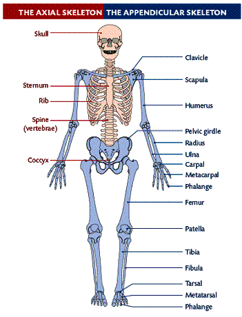
1. Axial Skeleton: skull, backbone, ribs and sternum.
2. Appendicular Skeleton: pectoral girdle, pelvic girdle and limbs.
Axial Skeleton
1. Skull: composed of 22 fused bones except for the mandible (lower jaw).
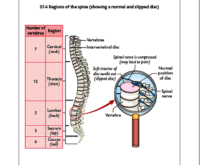
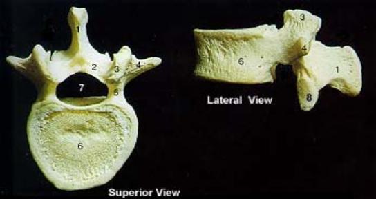
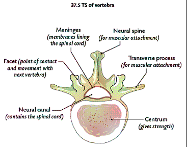
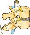
2. Spine or Backbone: Also known as the vertebral column. It is composed of 33 small bones in a line – cervical (7), thoracic (12), lumbar (5), sacrum (5), coccyx (4). The vertebrae of the sacrum and coccyx are fused together. The vertebrae of the other regions can move slightly giving flexibility to the backbone. There are cartilage discs between these that act as shock absorbers. They help to protect the vertebrae. The vertebrae are held in position by ligaments. Muscles attached to their surfaces also support the vertebrae. Spinal nerves emerge in pairs from the spinal cord between the vertebrae.
Rib Cage
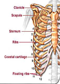
The rib cage consists of the sternum and 12 pairs of ribs. All 12 pairs all attached to the backbone. The first seven are true ribs joining also to the sternum. The next three are the false ribs, which also join to the seventh rib. The last two are the floating ribs. They are only joined at one end to the backbone. The sternum or breastbone is a flat thin bone at the centre of the chest wall.
The Appendicular Skeleton
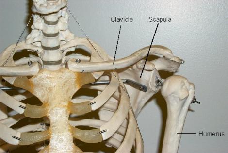
The Pectoral Girdle is composed of four bones – two clavicles (collar bones) and two scapulae (shoulder blades). The arms connect with the scapulae at a ball and socket joint.
The Pelvic Girdle
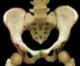
The Pelvic Girdle appears to be one large cylindrical bone but is actually six fused bones. The legs articulate with the pelvis at a ball and socket joint. The pelvis is securely attached to the backbone at the sacrum.
Limbs
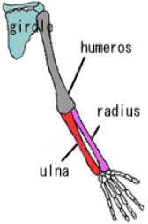
Human Forelimb (arm):
Human Hindlimb (leg):
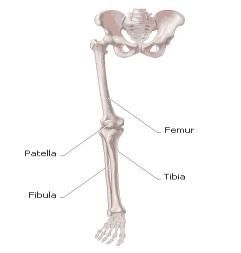
The arms and legs have similar design. There are long upper bones. In the arm it is the humerus and in the leg it is the femur. Also, there are two long bones in the medial region: The ulna and radius in the arm and the tibia and fibula in the leg.
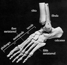
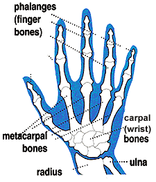
The foot and hand have similar bones. The carpals are in the wrist of the arm and the tarsals in the ankle of the leg. The metacarpals are in the palm of the hand and metatarsals in the hind foot. The phalanges are in the fingers and toes.
Long Bone Structure
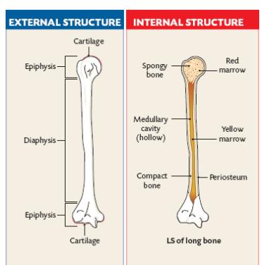
A membrane called the periosteum encloses long bones. It contains blood vessels and nerves.
The diaphysis is the shaft or long main portion of the bone. The epiphysis is at each end of the long bone and is formed separately of the diaphysis.
Cartilage is found at each end of the long bones. It is made of protein called collagen. Collagen fibres are wound within the surrounding material of calcium and phosphorous salts. The cartilage covers the epiphyses protecting them from friction and shock at freely moveable joints. Cartilage does not have blood vessels or nerves. Useful materials enter the cartilage by diffusion.
Types of Bones
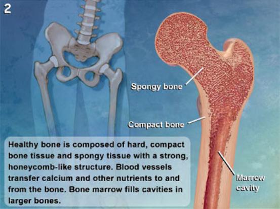
There are 2 types of bones: compact bone and spongy bone.
1. Compact Bone: Is a solid bone that at microscopic level has a concentric ring structure. It is made of bone cells called osteoblasts. These cells are growing within a material that is called a bone matrix. The bone matrix is made of 70% inorganic salts such as calcium phosphate and 30% protein called collagen. The calcium salts give the bone strength while the protein gives it flexibility. Blood vessels as well as nerve fibres are within the bone.
2. Spongy Bone: irregular openwork of thin plates of bone. It is also known as trabecular or cancellous bone. The mineral deposits are arranged as a system of struts. Bone marrow fills the spaces between the plates. The marrow cavity is the space within the diaphysis that also contains marrow.
Bone Marrow
There are 2 types of bone marrow:
1. Red Bone Marrow: Is responsible for the formation of red blood cells, white blood cells and platelets.
2. Yellow Bone Marrow: Is responsible for energy storage in the form of lipid (fat). This can be converted into red marrow if the body needs increased blood cell formation. It is only found in adults? medullary cavity.

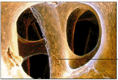
Inside view of cavities within spongy bone. They are filled with red bone marrow:
Bone Growth
The embryo is made of cartilage until about the 8th week of development. It then is replaced with bone. Osteoblasts produce the protein collagen. A hard covering of mainly calcium phosphate forms around the collagen. The osteoblasts become trapped within the calcium phosphate and become dormant bone cells.
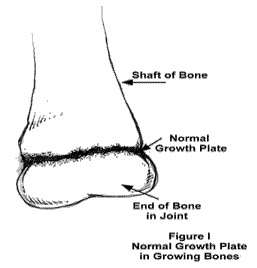
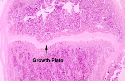
The bone becomes longer as a result of growth plates found between the diaphysis and the epiphysis. At this plate, cartilage is continuously formed and then turned into bone. This process is called ossification. This stops when a person reaches the adult age.
Bone Development
Even though bones stop growing in length in early adulthood, they can continue to increase in thickness or diameter throughout life in response to stress from increased muscle activity or to weight. The increase in diameter is called appositional growth. Osteoblasts in the periosteum form compact bone around the external bone surface. At the same time, osteoclasts in medullary cavity break down bone on the internal bone surface, around the medullary cavity. These two processes together increase the diameter of the bone and, at the same time, keep the bone from becoming excessively heavy and bulky.
Factors that affect bone growth:
1. Stress on the bones by physical activity. Osteoblasts are stimulated. More bone grows.
2. Lack of Stress causes bones to become thin.
3. Growth hormone and sex hormones increase bone size.
Joints
A joint is the junction between two or more bones. There are three major types of joints:
1. Fused Joints: These joints include the skull, sacrum, pelvis, and coccyx. As the name suggests, these joints are points where joints fuse or grow together. The place where they grow together is called the suture. These joints provide strength, support, and protection.
2. Slightly Moveable Joints: These joints are located between the vertebrae of the upper spine. There is cartilage within the joints. They help pad and protect the bones. The bones are held together by ligaments. The ligaments are tightly bound and limit the movement of the bones. This protects the spinal cord.
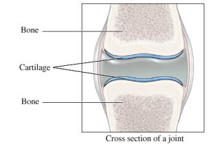
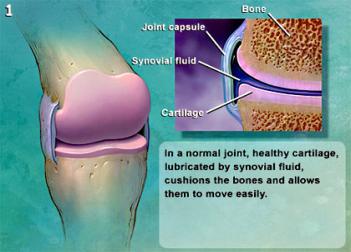
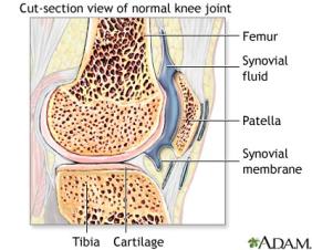
3. Freely Moveable or Synovial Joints: At these joints the ends of the bones are covered with cartilage and there is a cavity that separates the bones. The bones are held in place by ligaments which stop the bones from moving too much. In addition to the ligaments the two bones are joined together by sleeve-like capsule. The capsule encloses the synovial cavity. The outer layer of the capsule is composed of ligaments. As stated previously, the ligaments keep bones together preventing dislocation and control the range of movement. The inner layer of the capsule is the synovial membrane. The synovial membrane secretes the lubricating synovial fluid. Lubrication is essential to prevent frictional wear and tear. The cartilage at the contact ends of the bones also reduces friction. The cartilage pads also acts as shock absorbers against mechanical damage.
Classes of Synovial Joints
1. Gliding: The bones of these joints move across each other, back-and-forth and side-to-side. Examples are between the carpals of the wrist and tarsals of the ankle.
2. Pivot: These joints allow a turning movement. Examples are between the first and second vertebras when turning the head, between the ulna and the radius of the lower arm when turning the palm of the hand up or down.
3. Hinge: These joints allow movement in one plane during flexion and extension. They act, as the name implies, like the hinge of a door. Examples are bending the elbow or knee.
4. Ball and Socket: This type of joint permits movement in three planes, i.e., in all directions. Examples are the shoulder and hip joints.
Ligaments
Ligaments are strong, slightly elastic tissues that connect bone to bone at joints. These tissues are more flexible when warm. That is why you should gently warm up before exercising. Ligaments prevent dislocation of the joint and control the range of movement of the bones at the joint.
Tendons
Tendons are strong inelastic cords or bands of connective tissue that connect muscle to bone. They are composed of collagen and contain blood vessels. The inelastic tendon will not stretch when the muscle contracts. Therefore the full pull is transmitted to the bone and the full range of motion is accomplished.
Arthritis
There are 2 types of arthritis, Osteoarthritis and Rheumatoid Arthritis. Both of these conditions involve the swelling and inflammation of the joint. See your text book page 352 for more information on this topic.
Muscles
There are 3 types of muscle: skeletal, smooth, and cardiac.
Skeletal Muscle
Skeletal muscle, as its name implies, is the muscle attached to the skeleton. It is also called striated muscle. The contraction of skeletal muscle is under voluntary control. These muscles are mainly responsible for movement of the body. Other purposes are posture maintenance, support of the joints, and heat production. While its contraction is fast and strong, skeletal muscle tires easily.
Smooth Muscle
Smooth muscle is found in the walls of all the hollow organs of the body (except the heart). Its contraction reduces the size of these structures. Thus it regulates the flow of blood in the arteries, moves your breakfast along through your gastrointestinal tract, expels urine from your urinary bladder, sends babies out into the world from the uterus, and regulates the flow of air through the lungs. The contraction of smooth muscle is not under voluntary control. It is called involuntary muscle. It contracts slowly and is slow to tire.
Cardiac Muscle
Your heart is made of cardiac muscle. This type of muscle only exists in your heart. Unlike other types of muscle, cardiac muscle never gets tired. It works automatically and constantly without ever pausing to rest. Cardiac muscle contracts to squeeze blood out of your heart, and relaxes to fill your heart with blood.
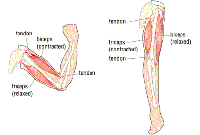
Antagonistic Skeletal Muscles
Antagonistic muscles are pairs of muscles. The action of one member is opposite to that of the other member. Muscles can contract but they do not have the ability to lengthen (stretch) themselves. They are arranged in pairs such that after one muscle or muscle group contracts, a skeleton transfers the movement to stretch another muscle or muscle group. The pairs of muscles that stretch each other are said to be antagonistic.
Example of an antagonistic muscle pair
The biceps and triceps muscles of the arm are an example of an antagonistic pair. Contraction of the biceps moves the arm toward the body and stretches the triceps. Contraction of the triceps extends the arm and stretches the biceps. In this example the bicep is said to be the flexor while the tricep is the extensor. Extensors are not as strong as flexors.